A neural atlas explorer
Find the evidence behind a spatial result.
NeuroViz brings datasets, atlas context, and provenance into one readable research surface—so every view can be examined, understood, and revisited.

Allen MERFISH-C57BL6J-638850
A spatial cell atlas built for questions that start with place.
The release contains 4.3 million QC-passed cells from one adult mouse brain, profiled with a 500-gene MERFISH panel across 59 coronal sections. Each cell can be considered in its anatomical, taxonomic, and registered spatial context.
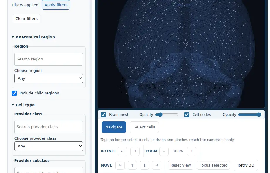
In the explorer
Move from a broad atlas to an inspectable cell cohort.
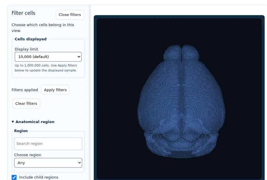
01 · Define a cohort
Filter cells without losing the anatomical question.
Start with an anatomical region, then refine the cohort by provider class or subclass. Gene-expression range, section, layer, and quality controls let you make the inclusion criteria explicit before interpreting a spatial pattern.
Open the Allen MERFISH filters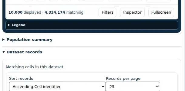
02 · Inspect the result set
Keep the cell data visible while you work.
The explorer reports both the displayed sample and the full matching count. Open the cell records to sort the current cohort and move from an aggregate spatial pattern to individual cell-level metadata, taxonomy, and measured expression.
View matching cell records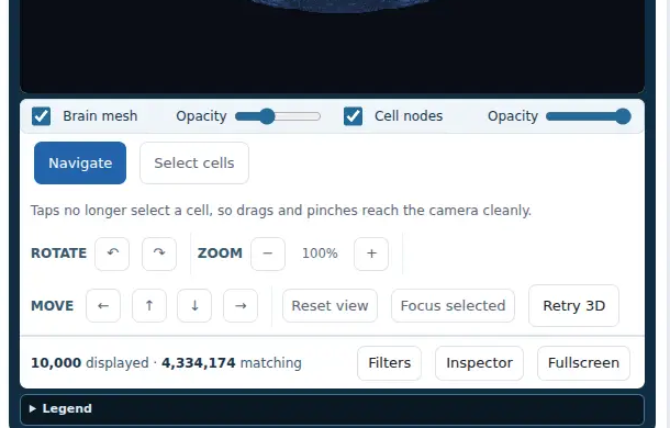
03 · Read the spatial context
Navigate the brain without leaving the cell view.
Rotate, zoom, pan, and reset the 3D camera to examine the selected cohort from a useful angle. Separate mesh and cell controls help reveal the spatial distribution while retaining the anatomical reference frame.
Explore the 3D cell view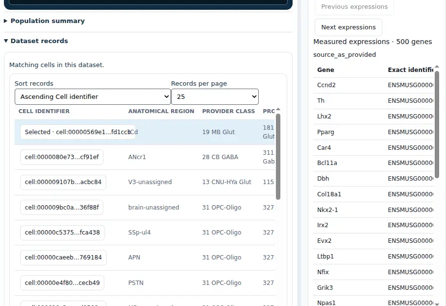
04 · Inspect an individual cell
Turn a selected cell into a reviewable observation.
Select a point in the brain or a row in the matching-cell table to open the cell inspector. Review its anatomical assignment, provider taxonomy, source section, and measured expression values without losing the cohort that led you there.
Open the cell inspectorResearch use cases
Start with a research question, not a visualization.
Explore cell identity and measured expression within 3D anatomy. These illustrative workflows make clear what the current explorer can support and where a proposed, validated extension is needed.
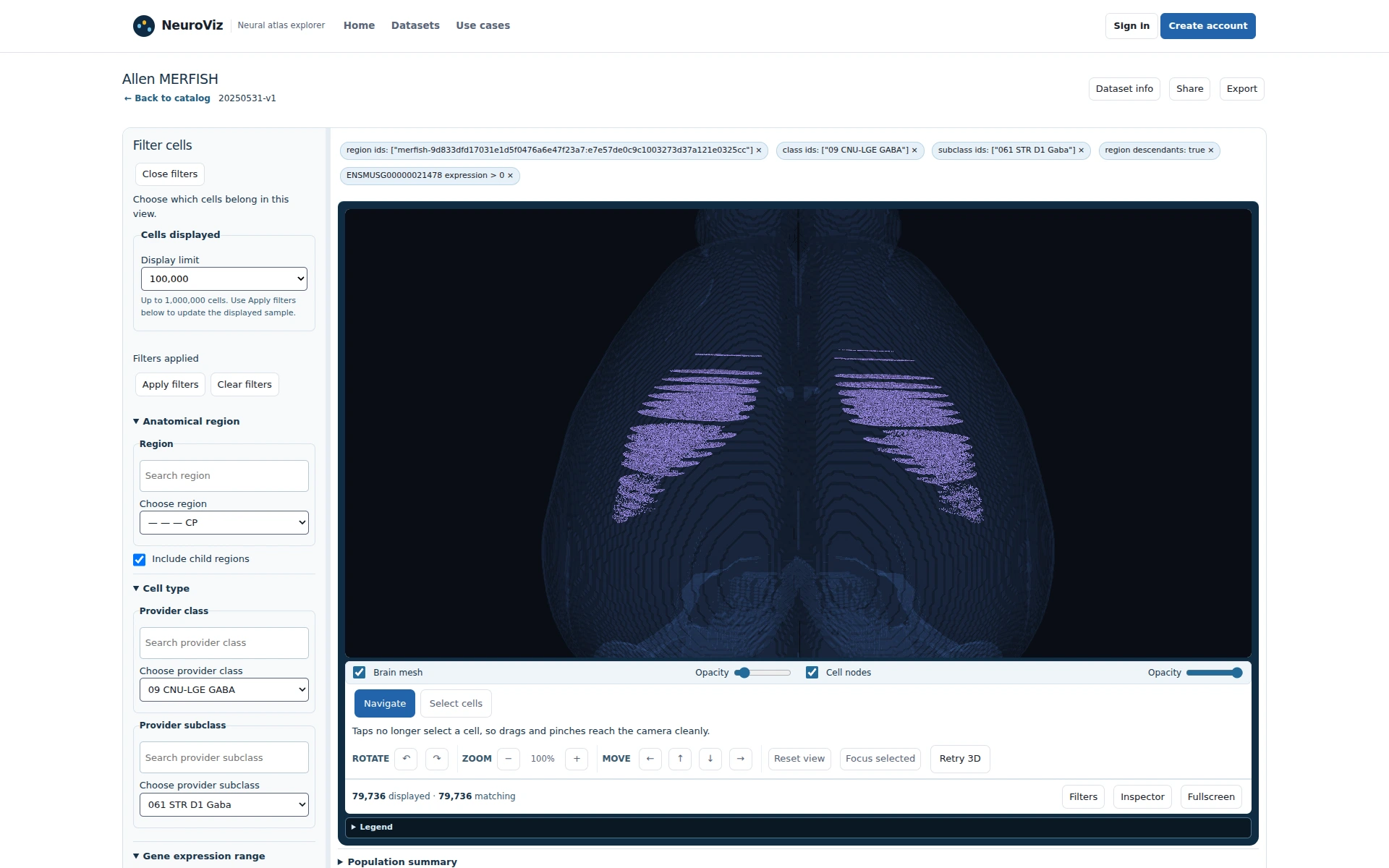 Explore nowMap D1 and D2 cells in the striatumHow do separately defined Drd1 and Drd2 cohorts map in CP?
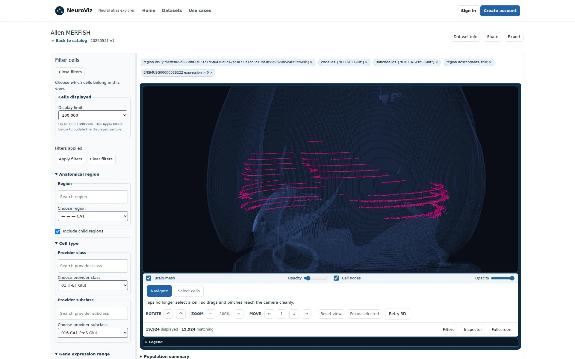 Explore nowExamine Calb1-positive cells in CA1What evidence is available for reviewing CA1-ProS Glut annotations?
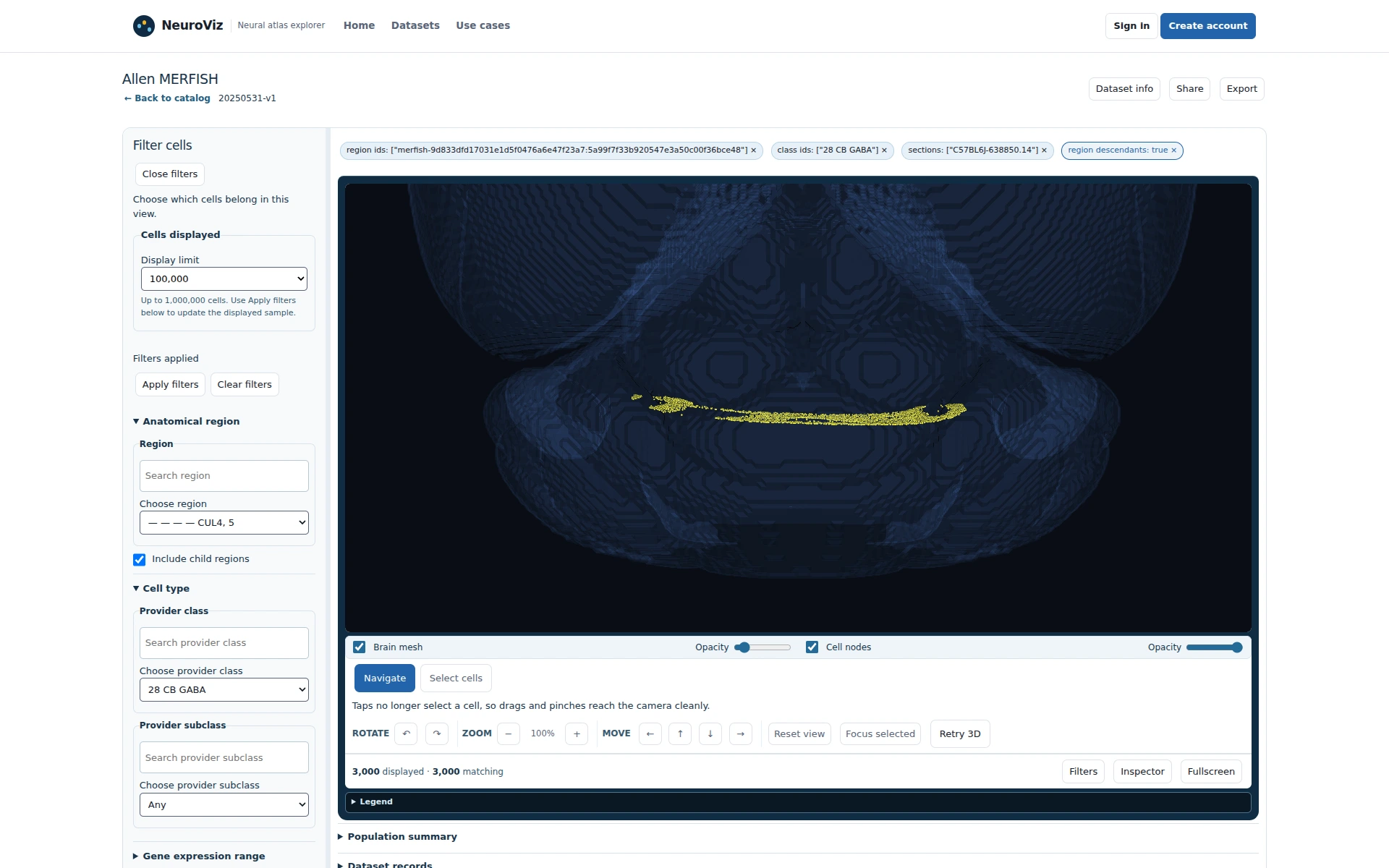 Explore nowCheck cerebellar continuity across sectionsWhat can adjacent source sections .13 and .14 show?
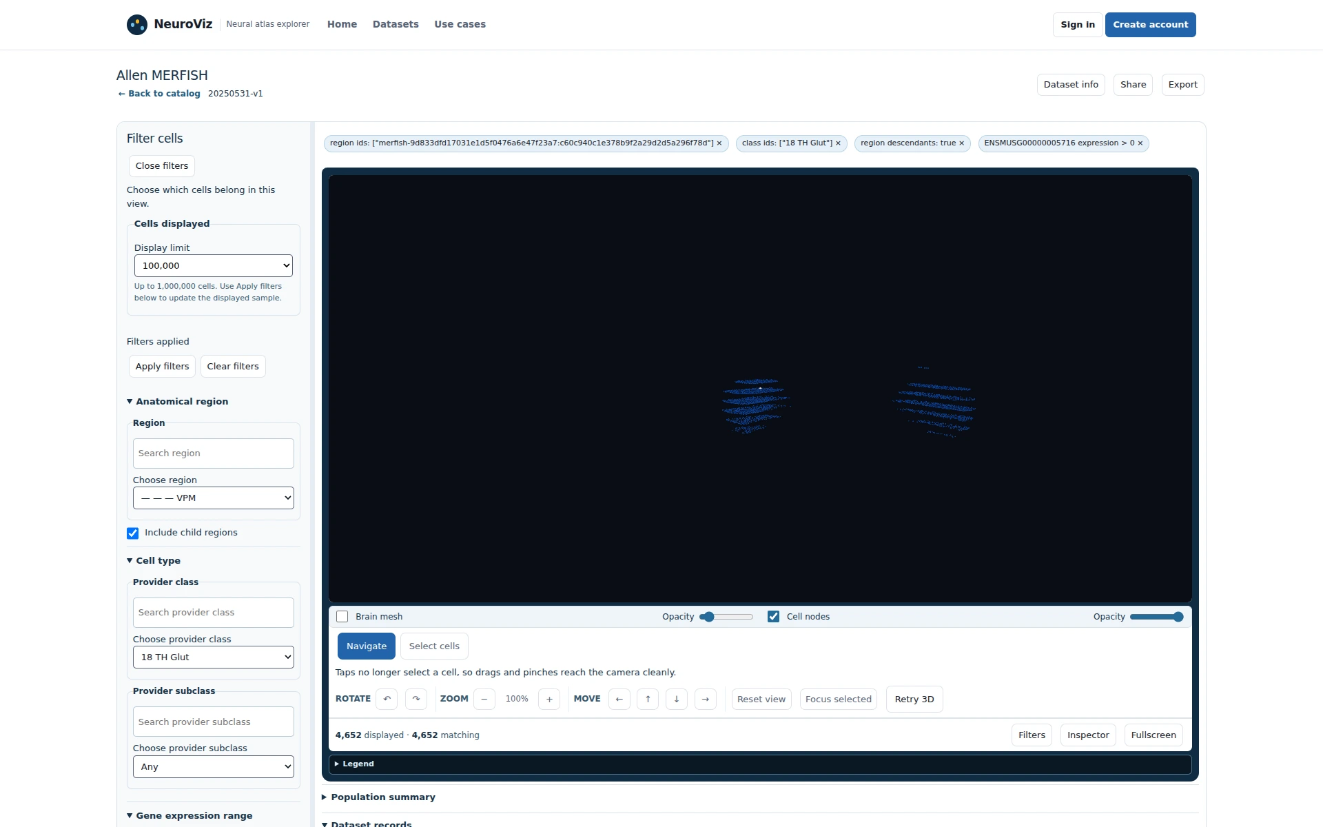 Explore nowExplore Pvalb-positive cells in the thalamusWhich VPM cohort should advance to spatial analysis?
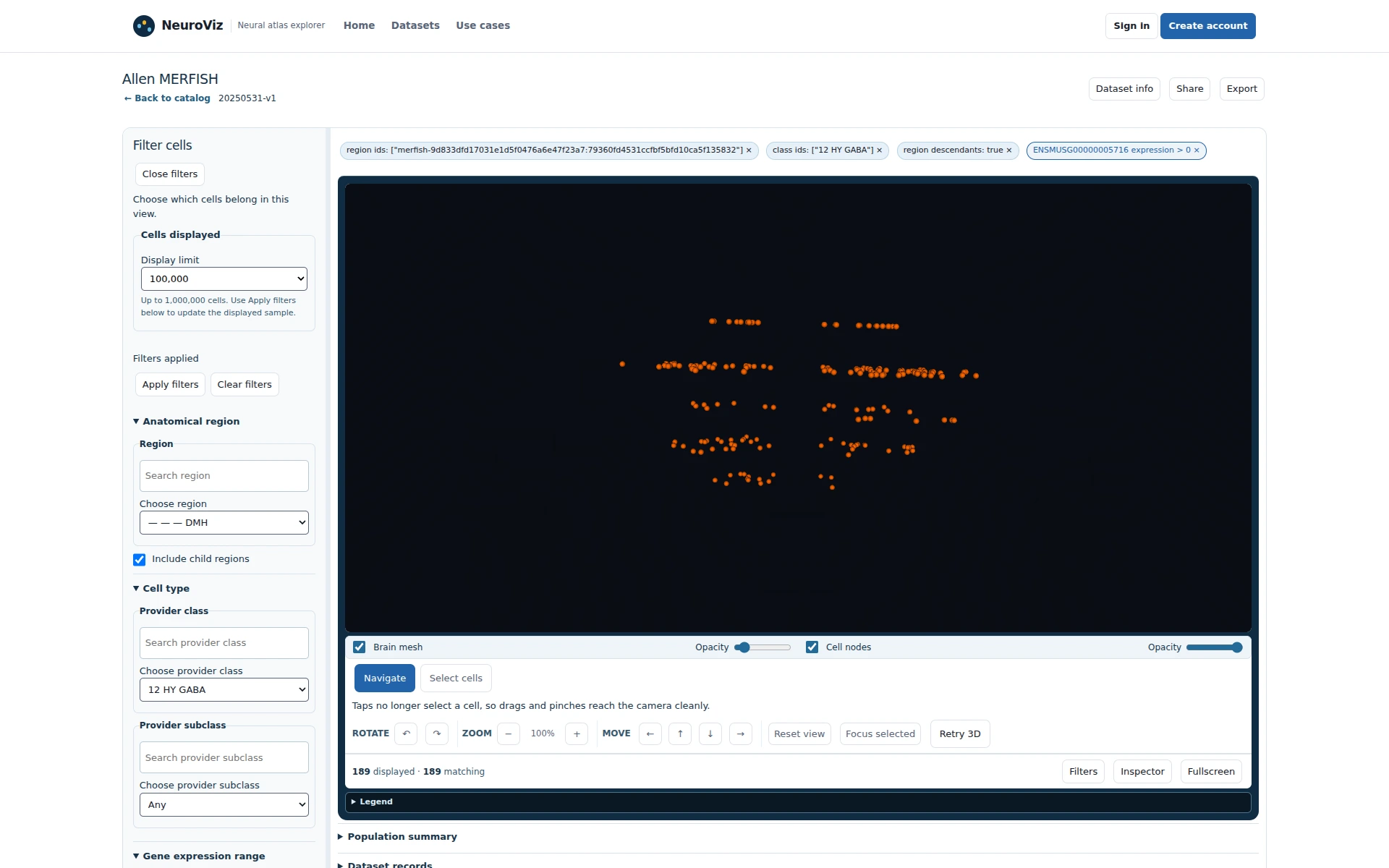 Explore nowPrepare a DMH reference cohortWhat can one public specimen contribute to a later study?
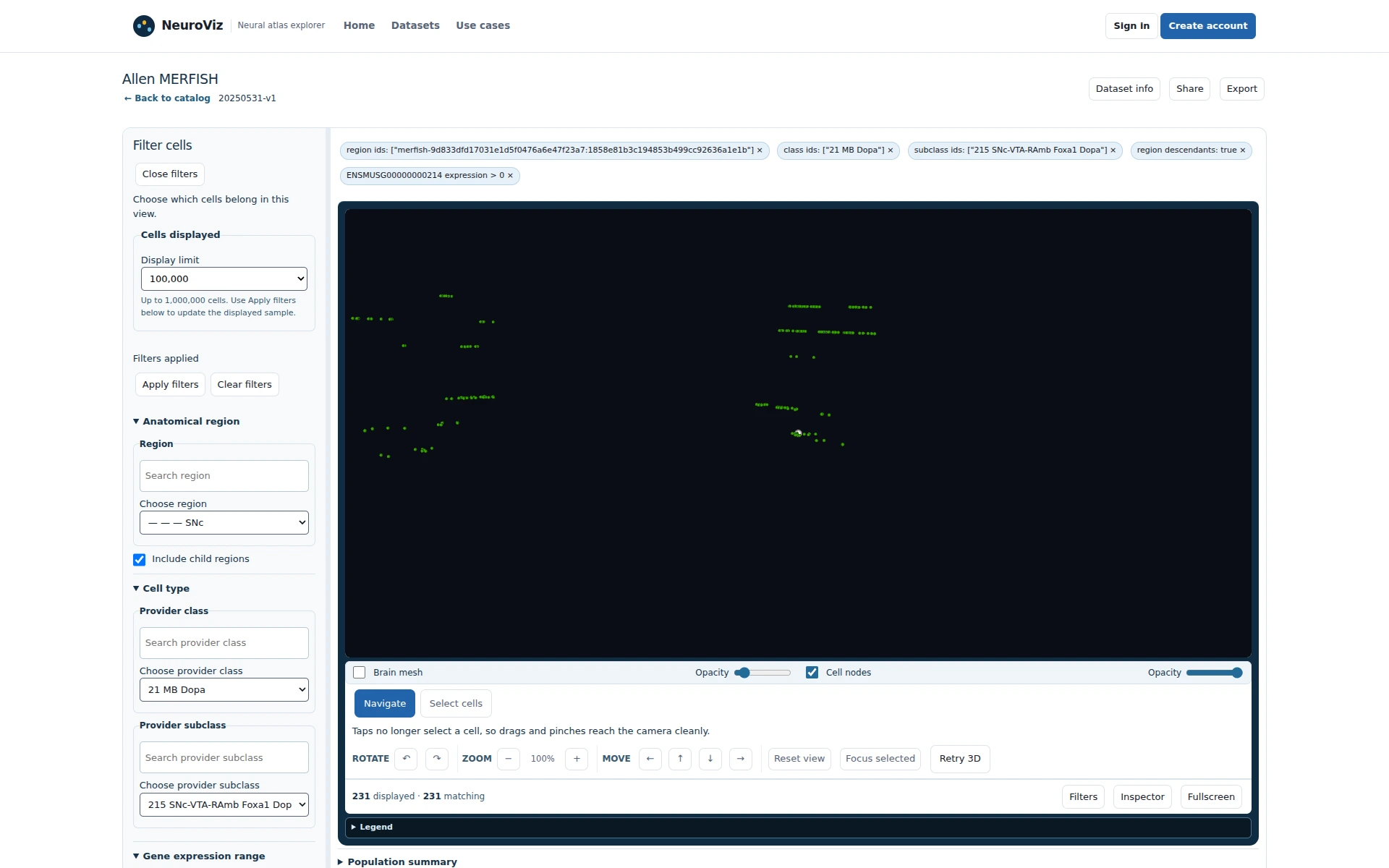 Explore nowReview midbrain candidates for follow-upWhich SNc cells and source sections merit a closer look?
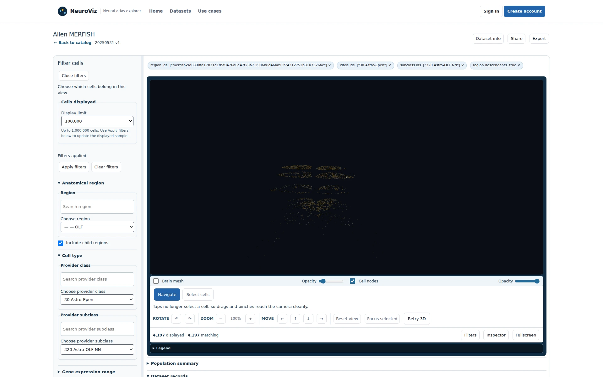 Explore nowExplore olfactory glial organizationWhich OLF glial cohort is ready for a later neighborhood analysis?